Red fright or green light? | A child with fever

babysimple-01-01

A 14 month year old boy has a fever for the past 4 days. The fever peaked at 38.5°C as measured at home. The child has been irritable and clingy since arriving at the GPs, but has not shown any obvious pain. There is no photophobia, neck stiffness or rash. There has been a non-productive cough for two days, which is worse at night. There is no history suggesting stridor, respiratory distress or wheeze. He has had a runny nose over this time. He is tolerating food and drink, although he is eating less than normal. He sometimes vomits after eating. There have been no changes to his bowel habits and he is producing about 6 wet nappies a day.

Examination is difficult. He cries at the slightest intrusion of his personal space by anyone other than his mother. His temperature is 37.2, although mum tells you he just had calpol about thirty minutes ago. His pulse is 120. There is subcostal and intercostal recession when he is crying, but none when the crying settles. His respiratory rate when he is settled is 40. His saturations are 96% on air. The anterior fontanelle is neither bulging nor sunken.

An ENT examination is almost impossible. You eventually find it to be normal. There is no rash top to toe. His chest sounds clear. His abdomen is soft.

You go to write up your findings to give yourself time to think whilst mum dresses the child. You have not found the source for the fever. You look at the PMH – he was a preterm baby at 34/40 weeks but has been generally well since. His development has been fine. All his vaccinations have been up to date. He is an only child, and no household contacts have been unwell.

But it’s only a ‘fever’ of 37.2 – that’s not significant

You only have a snapshot of the child’s temperature. You cannot discount the parental concerns about fever over the past four days based on a one-off reading just now. NICE state:

“Reported parental perception of a fever should be considered valid and taken seriously by healthcare professionals”

Then it’s a feverish child with no source! Admit, admit!

There are basically two things that mean a feverish child may need admission –a) suspicion of a serious bacterial infection or b) not coping with the illness itself e.g. dehydration, first ever/complex febrile convulsion, respiratory distress etc.

Regarding a) – the majority (>80-90%) of fevers in children will be self-limiting viral infections.

Regarding b) – the majority of children will cope absolutely fine with a febrile illness.

Our task is to identify the minority who have a problem with a) or b) for further assessment. We should advise the remainder to self-care at home with safety netting. Sometimes you cannot quite 100% pin down the source; this in itself does not necessarily require an urgent assessment.

How do you interpret the fever in light of the fact that the child took calpol?

It does not matter. The height of the fever in a child over 6 months does not predict a serious bacterial infection, nor does it tell you how the child is coping with the illness. The height of the fever is therefore not going to alter your management plan.

In infants less than 3 months, a fever greater than 38°C should be seen urgently by a paediatrician (red flag in NICE guidelines).

In infants between 3 and 6 months, a fever greater than 39°C is an amber flag in NICE guidelines.

Isn’t it great that NICE guidelines are perfect! We don’t have to think, just use the table!

The traffic light system is helpful. It does not replace clinical judgment. This reflects the fact it is difficult (?impossible) to create a rigid scoring system for something as complex and nuanced as assessing a feverish children. The most common bacterial infection the traffic light system misses is a UTI – in fact, about one fifth of the time a child with a UTI will score green. There has been a suggestion to add urinalysis to the NICE traffic light assessment of the feverish child (http://www.bmj.com/content/346/bmj.f866). However, there are also some who feel that this study considered only the traffic light system in isolation, without considering the NICE guideline’s holistically. The purpose of the traffic light system was not to diagnose but to help with the initial triage of feverish children. (http://www.bmj.com/content/348/bmj.g2056/rr/691345)

So what is this magical table?

Have a look: traffic light system for identifying risk of serious illness

The main thing to emphasise is that NICE states that if the child appears unwell to you, refer urgently. Your clinical gestalt is valuable, no matter your level of experience with children ( http://www.ncbi.nlm.nih.gov/pmc/articles/PMC3458229/).

What happened in this case?

It’s a fever of unknown source. There are some suggestions of a viral source (runny nose, non productive cough) but nothing definitive. There is no way you can predict what will happen in 4, 8 or 24 hours. The mother was told to contact us if the child remained clingy/crying frequently at home. Given the child had green flags at the time of assessment, and the mother knew when to come back or when to go to A&E, the child could be watched at home with appropriate safety nets.

Cool. Alternating paracetamol and tepid sponging then to keep T < 38.

NO! That is so 2008. You treat fever only so the child doesn’t feel distressed. You do not give antipyretics to target a temperature.

And you only give ibuprofen or paracetamol (at least initially). Alternating doses leads to frequent dosing errors. If the fever is not controlled with either agent alone, and the child is distressed, then maybe you could alternate them – but ask the parents to keep a written record of what they have given and when. And forget tepid sponging. Just dress the child in clothes which are appropriate for the room temperature.

Any specific advice for the parents?

NICE CKS mentions the following:


  • Look for signs of dehydration including poor urine output, dry mouth, sunken anterior fontanelle (usually closed by 18 months), absence of tears, sunken eyes, and poor overall appearance).
  • Offer regular fluids (if breastfeeding, continue this as normal) and encourage a higher intake if signs of dehydration develop.
  • Dress the child appropriately for their surroundings, with the aim of preventing overheating or shivering.
  • Avoid using tepid sponging (using cool water) to cool the child.
  • Check the child regularly, including during the night (how often depends on the situation).
  • Keep the child away from nursery or school while the fever persists, and notify the nursery or school of the illness.
  • Advise parents and carers to seek medical help if:
    • The child is getting dehydrated.
    • The child has a fit.
    • The child develops a non-blanching rash.
    • The fever lasts longer than 5 days.
    • The child is becoming more unwell.
    • They are distressed or concerned that they are unable to look after the child.


Early warning, late realisation

Early warning scores are in use throughout NHS hospitals to spot patients who are deteriorating. So where did they come from, and what are their limitations?

At Barts in 1997, it was noted that patients admitted to ITU from the wards had a high mortality.   During the summer and autumn of 1997, a new ‘patient at risk’ team (PART) was set up at Barts in Whitechapel. Their role was to assess ward based patients with a view to fixing things before they got worse, and thinking early about potential ITU/ceiling of care decisions.

Patients were selected for review by the PART based on a scoring system, where more deranged physiology led to a higher score. A certain score would trigger the nurse to call the ward doctor or the PART directly.

Noble stuff. Did it help? Of the 97 patients admitted to ITU over this six month period, 28 were seen by the PART within the preceding 48h. The incidence of CPR among this group pre-ITU admission was 3.6% compared to 30.4% among those patients not assessed by the PART (p<0.005). Mortality once at ITU was 25% for the PART group vs 44.9% for the non PART group, although this did not quite reach statistical signficiance (p=0.07).

Over time, various warning scores were developed, ranging from single parameters to aggregated scores. There is now a drive by the RCP to encourage NEWS, which is a nationalized early warning score.

What are the limitations? The National Patient Safety Agency published a report in 2007 which in part assessed how the early warning scores were working. 16 clinicians were interviewed, including 5 junior doctors. It’s worth reading this – key problems were communication, observations being seen as low priority and trigger fatigue (too many bleeps for “Just thought I’d let you know Mr Peters is scoring a 5…”)

From my own experience, there are three things I wish I understood better at the start of FY1 about using early warning scores.

1.    Communication

Personally, I sometimes spent more time trying to write a comprehensive and totally unambiguous management plan than actually with the sick patient. I was often confused when I then got bleeped asking what the plan was.

This was because I assumed that nurses will read the medical notes. It only dawned on me that nursing notes and medical notes are kept in separate folders about two months into my first placement. Lesson learnt: spending 30 seconds explaining the plan to the nurses saves you twenty minutes from having to revisit the patient when your plan wasn’t followed.

2.    Not all unwell patients score

There are some sick patients who don’t always score very highly on early warning scores. This includes patients with meningitis, patients with raised ICP, stroke, sepsis (especially neutropenic sepsis) and overdoses. It is worth mentioning that the elderly have a lower baseline temperature than the young, and that not only is the febrile response is blunted in 20-30% of elderly patients with acute infection, but a blunted response is associated with a poorer prognosis.

A patient’s change in their observations from their baseline is a better measure than the early warning score of how they are doing. A patient who is normally hypertensive with a BP drop from 175/100 to 100/65 in two hours doesn’t score on most systems, but this could lead to end organ hypoperfusion in this patient.

3.    Some not so unwell patients score

Lots of hospital inpatients will have a raised pulse from simply pain and anxiety. Many patients with well controlled COPD on home oxygen may score highly for oxygen and low sats on admission to hospital despite being at their baseline for these criteria.

Again, a patient’s change in their observations from their baseline is a better measure than the early warning score of how they are doing. A stable COPD patient whose sats have been between 88% and 92% for the past 2 days may move from 90% to 89% and suddenly score enough to trigger a colour change to amber, but this doesn’t mean there is a new acute problem.

What does this mean for the junior doctor on call?

Early warning scores are most effective at selecting a select group of patients that the nurses should generally seek a doctor’s opinion on. It may be end up being nothing serious; this is not a reason to get angry with the nurse. Your clinical assessment supersedes the score as the measure of the severity of the problem. For all subsequent dealings with this patient, you should use your knowledge of the situation together with the trend in observations to manage accordingly; you should not be using the early warning score alone to decide if the patient is improving or deteriorating.

Talking directly to the nurses after you have seen the patient will save you time in the long term. Unwell patients and the early warning score are far from perfectly correlated. Whilst it is true that for the whole group of hospital patients there is a correlation between early warning scores and general unwellness, there are specific disease processes that are not well matched to the early warning scores, and an individual’s variation from baseline supercedes the early warning score as a measure of how that individual patient is.

The early warning score is undoubtedly useful, but we need to remember it is a screening tool, nothing more.

Complete run through of the MRCP Part I – Day 2 of 40

Wow. This is a long syllabus. It’s taken me three hours to put this next section together, which leaves me with little time for pretty pictures/mnemonics. Let’s crack page 83.


Occupational asthma

This accounts for one sixth of all cases of asthma. Forget asbestosis, silicosis or smelly mould fancier’s lung – occupational asthma is the most common occupational lung disease.

Common allergens include flour, grain, wood dust, animal bits and isocyanates.

The history is more useful for excluding occupational asthma rather than confirming it i.e. if you have symptoms which are worse at home then at work, you don’t have occupational asthma. However, if your symptoms are worse at work, you may or may not have occupational asthma.

To be sure, you need to do confirm a relationship between asthma and work exposure by performing Serial measurements of PEFR at home and at work. Measurements should be made every two hours from waking to sleeping for four weeks, keeping treatment constant and documenting times at work.

Rhinitis and asthma often co-exist, including when occupational.

Once the relationship is confirmed, the patient must not work in that environment. At all.


Other common allergies

1. Latex allergy

Affects medical workers, people with spina bifida and rubber workers. The allergen is either natural or synthetic rubber

Remember the fruit latex syndrome in 30-50% of those with latex allergy. This involves allergy to avocado, banana, chestnut, kiwi, peach, tomato, potato and bell pepper.

2. Insect strings 

Usually bee and wasp stings

3. Drug allergy

I had no idea there was guidance specifically for drug allergy – see http://www.bsaci.org/guidelines/drug-allergy

MRCPwise, it’s probably worth going through the table of Type I – IV hypersensitivities on page 45. The guidance tells you about risk factors for developing allergies as well as how to test for allergy semi-safely.

I also learnt from this guidance that intradermal tests are more sensitive but less specific than skin patch tests, and have a higher risk of causing anaphylaxis.

4. Food allergy

The Medical Masterclass focuses on nut allergies, which are the commonest food allergies. Peanuts account for >50% of all food allergies.

In children, failure to thrive, vomiting and eczema can all be explained by food allergy.

Distinguishing between intolerance and allergy can be tricky. Allergy happens within seconds of ingestion, and only minute quantities are needed. In contrast, intolerances take hours to develop, and a small quantity poses no problem.

Intolerance e.g. lactose intolerance produces bloating and diarrhoea. Allergies may provoke eczema, reflux, GORD as well as anaphylaxis.

A food diary, combined with skin prick testing/appropriate IgE tests can be helpful.


Angioedema and urticaria

I found the patient information to the British Association of Dermatologists helpful:

The weals of urticaria may be flesh-coloured, pink or red. They can be of different shapes and sizes, but usually look like nettle stings. An important feature of urticaria is that although the rash may persist for weeks, individual lesions usually disappear within a day, and often last only a matter of hours. However, they sometimes leave bruising especially in children. New weals may then appear in other areas. In ordinary urticaria, the weals can occur anywhere on the body, at any time.

The deeper swellings of angioedema occur most frequently on the eyelids, lips and sometimes in the mouth, but they may occur anywhere. They are not usually itchy, and tend to settle within a few days. If the hands and feet are affected, they may feel tight and painful.

Angioedema occurs beneath the dermis and affects the skin and mucosa, whereas urticarial occurs above the dermis and only affects the skin. Angioedema is painful and tense; urticarial is itchy.

Almost any medicine can cause ‘acute’ urticaria, but painkillers (especially aspirin and medicines like ibuprofen), antibiotics (especially penicillins) and vaccinations are most likely to be responsible. Angioedema, in particular, can be caused by a type of drug (ACE inhibitors) used to treat high blood pressure.

Where does C1 esterase deficiency come into it?

Hereditary angioedema causes provoked or unprovoked attacks lasting 2-5 days of laryngeal odema, subcutaneous swelling and abdominal pain. The tests you want initially is C4 level and if this is low, then C1 INH function and level of protein.

What about ACE inhibitors?

Any individual can get angioedema from ACE inhibitors, though it is more common in Afro-Carribeans. It can occur years after uneventful ACE inhibitor use, and is poorly responsive to antihistamines, corticosteroids or adrenaline. The episodes may persist for some months after stopping the ACE inhibitor. Switching to ARBs is usually successful.

What is the management of angioedema?

See http://www.patient.co.uk/doctor/angio-oedema. In general, the acute attack usually needs nothing, although if there is airway compromise then manage as anaphylaxis. Chronic angioedema should be started with an antihistamine, although antihistamines don’t always work in angioedema.

And urticaria? 

First line is a non sedatating H1 antihistamine e.g. cetirizine plus three days of 50mg prednisolone.


Allergen sensitisation…

Step 1: Choose an allergen

Step 2: Get a B-cell to get excited by this allergen

Step 3: Invite this B-cell to mature into a plasma cell and secrete IgE specific to this antigen

Step 4: Stick the IgEs onto mast cells

You have now sensitized the patient!

Primary prevention of allergy prevents this sensitization from happening. This means allergen avoidance.

Secondary avoidance means preventing further sensitization once the process has started. This means leaving the job which triggers occupational asthma.

…and immunotherapy

Indications:

  1. Hay fever that cannot be controlled with the usual management (unless asthmatic)
    To manage hay fever needs allergen avoidance, education and meds. Avoidance needs knowledge of the allergen, which can be found by skin prick tests
    Topical antihistamines for nasal symptoms are great, but only tackle nasal symptoms. Topical intranasal steroids are safe long term and more effective, but risk dryness, crusting and bleeding.
    Oral antihistamines are the mainstay, and work on neutrally mediated itch, sneezing and rhinorrhea. They don’t cover the blocked nose as well as intranasal steroids however.
    There are also nasal decongestants, and eye drops eg sodium cromoglicate

  2. Hypersensitivity to wasp and bee venom

What happens?

Administer gradually increasing quantities of specific allergens to patients with IgE-mediated conditions until a dose is reached that is effective in reducing disease severity from natural exposure.

How does it work?

Eventually, the allergen stimulates IgG rather than IgE production.

Contraindications?

ACE inhibitors – they may trigger an anaphlyactoid reaction (anaphylactoid is basically anaphylaxis that is not IgE mediated)

Beta blockers – reduce the efficacy of adrenaline, which may be needed


Allergen avoidance

The main allergens are house dust mite, mould, pollen, pets and dust. Advice on avoidance is given at http://www.allergyuk.org/the-management-of-allergy/allergen-avoidance


Natural history of allergic diseases

Asthma stops in about 55% of children by the time they reach adulthood. This is more likely the earlier the asthma started.

Eczema stops in around a third of children when they reach adulthood.

Rhinitis tends to persist into adulthood.

There is a concept called the ‘allergic march’ – see http://www.worldallergy.org/professional/allergic_diseases_center/allergic_march/


The tests

The skin prick test needs resuscitation facilities to hand, but is most useful for food allergy and rhinitis (include tree pollen, grass and weed)

Not great if: dark skin, dermatitis, dermatographism, unable to stop antihistamines for 48 hours before

Allergens introduced to skin through pricking. If positive, a wheal of at least 2/3mm develops. Negative predictive value is 95%, but positive predictive value is only 50%, which means a good history makes a big difference.

A skin test for A. fumigatus is done for ABPA, as well as total IgE.

RAST and ELISA are blood tests looking for specific IgEs. They are expensive, but are not affected by antihistamines or the condition of the skin.

Oral food challenge is the definitive but risky test for food allergy.

Patch testing – read at 48-72 hours, and is used for contact dermatitis and Type IV stuff in general.

Lactose intolerance is tested by the Lactose hydrogen breath test:

The patient ingests a standardised amount of lactose (2 g/kg, maximum 25 g) after an overnight fast with measurement of exhaled hydrogen over a 2- to 3-hour period. A positive test is an increase of >20 ppm of exhaled hydrogen after approximately 60 minutes. (BMJ Learning)

Why we should never ask if your saddle-region is numb

I guess the ideal doctor would have experienced first hand every condition that can affect a human. He or she would then be able to pattern recognise what the patient is describing, and tease out the relevant information during history taking to diagnose the problem.

The ideal doctor is not possible, so we instead rely on reading textbooks, reviews and listening to clinical pearls from experienced colleagues. We also gain experience over time.

The common theme to all of these sources is that ultimately the information comes from patients. Patients are our ultimate clinical tutor for history taking.

This is what makes this podcast on back pain especially brilliant. You have the uniquely useful situation where the patient is the doctor, describing his own case.

I listened to this podcast during my car journeys, and one point stuck with me. When the doctor-patient was asked “Do you have numbness around your buttocks?” he would always answer no. When instead he was asked “Does it feel different when you sit down?” he would answer yes. He found it difficult to explain exactly how it was different, but it just felt funny.

He ended up having cauda equina.

So, with this in mind here’s the case. A 65-year-old man had a background of chronic back pain. Whilst bending over to do some gardening, he had a sudden onset of pain in his lumbar back.

The pain was central and 10/10 severity. It worsened with sudden movements, coughing and weight bearing. It was relieved by lying flat. The pain stopped him sleeping, and was progressively worsening. There was radiation to the right leg only. There was some tingling in the left leg. He was finding it harder to walk, and he was normally an independently mobile man.

A red flag review revealed:

Fracture risk:

No known osteoporosis
Osteoporosis risk factors: maternal hip fracture
No major or minor trauma

Malignancy risk:

PMH of melanomas, surgically excised 5 years ago
No weight loss
Some night sweats for the past week with fever for the past two days
No thoracic pain
No point tenderness
Age > 50

Infection risk:

No immunosuppression
Fevers and night sweats as noted
Age > 50

Neurological involvement, including CE:

“Does it feel different when you sit down?”
“Yes”
“How so?”
“It feels like I’m always sitting on a pillow.”

“Any changes to your bowel habits?”
“I haven’t gone in the past two days.”
“And does it feel different in any way when you wipe yourself?”
“Yes – it’s numb.”

No bladder symptoms
No weakness
Some new paraesthesia in the left foot

On examination

There was diffuse erythema and warmth around the L3/L4 area. No point tenderness, but tender to palpation diffusely from L1-L5 ish. The pain was worst centrally, and there was no paraspinal tenderness or obvious muscle spasm.

Straight leg raise caused pain at 5 degrees elevation, with pain spreading all the way down the leg into the feet as well as pain in the back. This was bilateral. Ankle dorsiflexion worsened the pain, again bilaterally.

Neurologically, power was intact. All lower limb reflexes were normal, with downgoing plantar reflexes. Sensation was abnormal perianally. The patient described it as lots of layers of cloth between the skin and the stimulus.

I organised an urgent MRI and referred to orthopaedics. I’ll post tomorrow what the outcome of all this was.

The case bore striking resemblance to the podcast’s story, of ‘different’ but not numb perianal sensation. On a BMJ article on cauda equina, it gives a patient’s perspective which reinforces this further:

“…I remember wiping myself with the toilet paper and it feeling decidedly odd—not completely numb but distant. It was my refusal to admit to numbness that fooled my general practitioner. He asked if I could feel him touching me, not whether his touch felt normal…”

I guess the lesson from this post is: don’t just ask for the presence or absence of sensation. Ask the patient to describe if the sensation is different in any way. This goes not just for cauda equina, but history taking and examination for sensation in general.


An n=1 trial into how a patient’s drawings reveal his underlying mood and maybe treat his depression

A man with vascular dementia was admitted following a suicide attempt. The Crisis consultant felt it was high risk, and recommended him to be admitted.

There’s “oh, dementia” and “oh…dementia”. This patient belonged in the first category. He looked after his house alone, and kept it in immaculate condition. He self-cared better than most students.  He had the air of an aged rock star, looking back over his life with pride and no regrets yet probably quite looking forward to living in a gentler gear.  The only time his dementia surfaced in our conversation was when he would freeze mid sentence, suddenly unaware of what he was talking about and as much embarrassed as frustrated by this.

I noticed how he used tricks to work around his memory problems which were quite similar to the memory tricks I use for learning medicine.  If he could not remember the name of someone, he would find a picture to remind him of them, and flood the picture with as many details as he could which would trigger off his memory.

When I can’t remember the features of a medical problem, I try and create an image to link them all together. I think that the reason this works so well is not so much the image, but that the very process of creating them forces you to engage with the material that much more.  Have a look at this totally not peer reviewed summary.

It seems this man was self-administering cognitive rehabilitation. Cognitive rehabilitation is defined as “any intervention strategy or technique which intends to enable clients or patients, and their families, to live with, manage, by-pass, reduce or come to terms with deficits precipitated by injury to the brain.” Examples of cognitive rehabilitation strategies include:

  • Build on the memory skills the patient already has using prompts to achieve whatever you need. Then fade the prompts. This video on errorless learning demonstrates it really well.
  • Use alternative skills to compensate e.g. pictures of clothes to remind you what the washing machine is used for.

This patient enjoyed drawing throughout his life. It seems he was using his presumably well developed visual neural circuitry to compensate for his short term declarative memory problems.

He wanted to show me his sketches. The first one was of Elvis Presley. It was quite impressive, especially given he did it at 3am.

Done at 3am. Quite impressive for an inpatient on a dementia ward.

Then he asked me something which caught me off guard:

“Can you see the anger?”

I looked at the drawing with my anger-filter switched on to maximum.

“His expression?” I suggested initially pointing to the face, and then further outwards looking optimistically for any signs of agreement.

“You’re nearly there. It’s all these lines.”

He pointed to the strokes towards the edge of the drawing.

“You see these lines? They are much darker. When someone makes me angry, or I’m getting frustrated, I don’t draw delicate lines. Can you see the anger in those lines?”

The angry bits are highlighted in red. Note how the lines are darker.

I could. It’s how I feel with Adobe Illustrator and the Image Trace function has been butchering my sketches. Sometimes I just give up and make the whole thing MS Paint-y. That’s a sign I got angry.

“Every time I’m angry, I remember it in my drawings. I remember what made me make those lines darker. It’s usually a loud noise, or someone disturbing me.”

I then realised his drawings might be one of the best ways of assessing his mood. This patient was giving us something that most advanced brain scan could never pick up. It was actually a pretty good functional assessment of his mood, recorded as is and without any attempt to pigeon hole his thoughts and feelings into any questionnaire criteria.

I looked at his old sketches with him. He could tell me exactly what he was feeling at the time, where he was and what he was thinking when he drew them. You could argue we do not know that he was not confabulating for the old sketches. However, the events he described surrounding the recent sketches done whilst an inpatient had actions associated with them that were verifiable with other members of staff e.g. a patient walking into him one evening. This is a man who was normally unable to recall events of the day before. It seems his drawings allow him to access memories of the recent past by coding emotionally significant events into their very structure.

Could this skill be used for his benefit? One of his greatest frustrations was not knowing what happened in the ‘in between time’. This was the time between the last few hours and the olden days, such as one week ago. Not knowing what happened depresses him. I could see why, given how well his other cognitive functions were and his high level of insight.

I wanted to see if we could help him help himself. I suggested keeping a drawing diary, and that when significant events happen that he wants to remember, perhaps he could sketch whatever he feels would capture it best. Although this would not capture all the details such as what he had for breakfast three days ago, it would capture the most significant memories. If we could capture those memories, I felt he would be less depressed.

He has decided to give it a go. He is also on mirtazapine 30mg NOCTE.

14/5/13: He has improved considerably, and his newer drawings reflect this.

4 doctors, 4 completely different management plans

salt-feature

An man in his early 80s with a background of Type I diabetes is day 1 post above knee amputation.

You are called to see him at 5pm because of a dropping urine output. He has had no urine output at all for the past 4 hours. In the 12 hours before this, he has passed 180ml of urine in total. He weighs 63.5kg.

On examination, he is haemodynamically stable (BP 130/80, pulse 80). His mucous membranes are a little dry, but you also notice pitting ankle edema in his remaining right leg. The JVP is not visible and his chest is clear. He is a bit more drowsy than when you saw him on the ward round that morning.

You flush his catheter, but this does not make a difference.

Of note, his Hb was noted earlier to be 7.6 g/dL, and he has been receiving a unit of blood at a 3 hourly rate for the past 2 hours, with another unit due immediately afterwards. He has not been eating post operatively, and so has been put on an insulin sliding scale, receiving 1 litre of dextrose every 10 hours for the past 5 hours. Before this, he was on Hartmann’s every 10 hours since the operation.

You also note his sodium to be 116 that morning. This was down from 125 the day before, and from 135 a few days before the operation.  His eGFR was seemingly better than ever, at >90mls/min/1.73m2 compared to 75 90mls/min/1.73m2 prior to surgery.

Medications wise, he was normally on insulin, furosemide 40mg, and ramipril 2.5 OD. He was not on any other heart failure medications (in particular, no beta blockers) and had no documented PMH of heart failure. He had been started on paracetamol 1g QDS, gabapentin 300mg OD (due to increase the next day to 300mg BD, then 300mg TDS), diclofenac 50mg TDS and PRN oramorph.

He had also started to feel a bit nauseous for the past hour, but had not vomited anything (BM 11.1, capillary ketones 0.1).

What do you make of the fluid used for the sliding scale?

The term ‘variable rate intravenous insulin infusion’ (VRIII) should replace the ambiguous term ‘sliding scale’.

Sorry, what do you make of the fluid used for the VRIII?

5% dextrose is not recommended as the fluid of choice for a VRIII. It is associated with hyponatremia. The first line fluid for a VRIII is 0.45% saline with 5% glucose and 0.15% KCl.

Fluid Drug added Dose Volume Route Rate
0.45% Sodium Chloride with 5% Dextrose Potassium Chloride 20 mmol 1 litre IV 12 hours

Huh? I don’t think my hospital even has that.

They probably don’t. It’s about three times as expensive as plain old 5% dextrose. Once there is greater appreciation of the guidelines published by Diabetes UK it should be ordered more, leading to a drop in price.

 So what do I use in the meantime?

If you can get two cannulas in, you can give 10% glucose with 0.15% potassium chloride at 60 ml/hr with a continuous VRIII in one cannula, and 0.9% saline at 60 ml/hr in the other.

Fluid Drug added Dose Volume Route Rate
0.9% Sodium Chloride 1 litre IV 18 hours
10% Dextrose Potassium Chloride 20 mmol 1 litre IV 18 hours

If you can not get two cannulas in and you have lovely nursing staff, you can switch between 10% dextrose with 0.15% KCL with a continuous VRIII if the capillary blood glucose is 14 mmol/L or less and 0.9% saline with 0.15% KCl if the capillary blood glucose is 15 mmol/L or more.  This does mean the nursing staff will have to change the bags each time the glucose level crosses 15, and fluid balance charts will be harder to accurately complete.

Fluid Drug added Dose Volume Route Rate
0.9% Sodium Chlorideif capillary glucose 15 mmol/L or greater Potassium
Chloride
20 mmol 1 litre IV 10 hours
10% Dextroseif capillary glucose 14 mmol/L or less Potassium Chloride 20 mmol 1 litre IV 10 hours

If you cannot get two cannulas in and you do not think switching the fluid bag is practical, at least give 0.18% saline with 4% glucose with 0.15% potassium chloride rather than 5% dextrose with a VRIII. This is however still associated with hyponatremia and not recommended for paediatric VRIIIs.

Fluid Drug added Dose Volume Route Rate
0.18% Sodium Chloride with 4% Dextrose Potassium
Chloride
20mmol 1 litre IV 10 hours

When exactly do surgical patients go on a VRIII? 

In general, when diabetic patients who have insulin requirements miss more than one meal they will go on a VRIII. The threshold for starting a VRIII is reduced when the patient is dependent on insulin (Type I and severe Type II) and when the patient has poorly controlled blood glucose.

Should we stop all other insulins before starting a VRIII?

No. Stop all insulins apart from any long acting insulin analogues e.g. Lantus.

What do you make of the sudden improvement in his kidney function?

How do we know there is actually an improvement in his kidney function? eGFR is calculated using the modified MDRD equation. The MDRD equation takes into account age, sex and whether or not the patient is black. These variables are used because they relate to total muscle mass, which in turns determines serum creatinine. If you visit renal.org’s guidance on the eGFR, you need to be cautious about interpreting it in patients of extreme body types. This is because the total muscle mass of these people may be different to what you would expect for their age/sex/race. Renal.org specifically cautions about using eGFR in amputees.

This patient could very well be going into AKI despite a reduction in his creatinine.

Why was he hyponatremic?

The causes of hyponatremia can be split into hypovolemic, euvolemic and hypervolemic. There’s another blog post on hyponatremia.

The release of ADH is either appropriate or inappropriate. It is appropriate when the serum osmolality is high, and when the effective circulating volume is low. If these conditions are not met, then ADH secretion is inappropriate.

Any cause of a decreased effective circulating volume eventually stimulates ADH. This is appropriate from the point of view of the circulation, but a bit dumb from the point of view of the overall total body fluid and its distribution. However, the stretch receptors in arteries and the heart don’t know the body’s total fluid status. This is why fluid overload states, like heart failure, cirrhosis and renal failure which have a reduced effective circulating volume but increased total body fluid, will tend to become hyponatremic.

It makes sense then to treat this with fluid restriction. However, there was a tailing urine output. Would you really fluid restrict someone with a gradually decreasing urine output?

I did an ABG to get a quick recent Na level, as well as look at his pH and potassium in case this really was AKI.

pH 7.44

pCO2 5.4 kPa

pO2 12.1 kPa on room air

BE +3.4

Na 114 mmol/L

K 4.5 mmol/L

Lactate 1.3 mmol/L

There were no neurological signs of hyponatremia. I discussed this case with the renal registrar, who said:

“It’s probably dilutional hyponatremia. Give him the second unit of blood. What fluid were you using on the sliding scale? Watch carefully for signs of overload. Give 40mg furosemide. Treat any low blood glucose with non fluids if possible. I know the patient is hyponatremic, but furosemide will probably raise the sodium in this case. I know the urine output is dropping, but post operatively this can happen without being in AKI. Don’t use the urine output to guide fluid input at the moment.”

Impossible potassium

The prize for the most extreme hypokalemia ever is not awarded solely for the magnitude of the hypokalemia. It is much more impressive when the potassium refuses to rise above 2.5 despite more than 160mmol of potassium being given per day for 5 days, as in the case of a lady in her mid 80s who had come in for an EVAR.

Her operation was uneventful. Day 1 post operatively, her potassium sank to 2.6 and never rose above this value till the day this post was written some 6 days later.

Why?

The causes of any serum electrolyte depletion can be split into inadequate intake, intracellular shift and excess losses.

Our patient was getting about 160mmol of potassium a day. Her intake was clearly more than sufficient.

Intracellular shift of potassium is usually caused by one of three things:

  • insulin (as in refeeding syndrome, the best condition ever)
  • a beta-adrenoreceptor agonist like salbutamol
  • alkalosis

Our patient was having some occasional salbutamol for recently feeling SOB, but nowhere near enough to cause K of 2.2 as it was yesterday morning. A venous bicarbonate was 23.

So she must have been she losing it from somewhere…

GI fluid is potassium rich, but post operatively there had been no diarrhoea or vomiting. A CT Abdomen showed no anastomic leak. She was on no laxitives.

What about urine?

She was on no diuretics. That said, her magnesium was 0.4-0.5 since the operation. Her electrolytes seemed to worsen when she had her fortisips resumed. I was thinking of refeeding syndrome, which as you may be starting to realise, is fast becoming my favourite thing ever (her phosphate was low-normal).

The team initially wanted to treat her magnesium cautiously, with oral supplements. However, the magnesium barely budged on this for 5 days. In fact, reading the BNF will reveal that oral magnesium is only for preventing recurrence of the deficit, not for correction of a deficit. I decided to switch to IV magnesium after 5 days of this, on the grounds that hypomagnesemia can make hypokalemia impossible to correct. I also discussed the case with the endocrine registrar, who suggested adding urine U&Es to determine if the kidneys are to blame.

Why exactly does a lack of magnesium lead to persistently low potassium?

See this review. The gist is that there are magnesium-inhibited ROMK channels in the late distal tubule/collecting duct that let potassium into the urine. If you lose the magnesium, those channels go into freeflow mode.

What were the results of the urine U&Es, and how did you interperate them?

The fractional Na excretion was about 1.2%, which is normal for a euvolemic patient. <1% would go with pre-renal failure, or a normal response to hypovolemia as the kidneys try to preserve sodium. Anthing above 2% is suggestive of a problem with sodium reabsorption, and as most reabsorption happens in the tubules this suggests a tubular problem, like acute tubular necrosis.

The normal bicarbonate pushes us away from renal tubular acidosis as a cause for the hypokalemia, although does not rule it out.

So what’s your conclusion?

In our patient’s case, the amount of potassium lost was inappropriate for the serum level of potassium, so this suggested the cause of the hypokalemia was renal. In light of the generally OK function of the tubules as suggested by the fractional Na excretion, and the lack of acidosis as suggested by the bicarbonate (though I do appreciate a normal bicarbonate is not proof of normal pH), this was not a tubular problem, and in particular not renal tubular acidosis. Something was selectively letting potassium leak through the kidneys.

My money is on low magnesium helping K to leak via the ROMK channel. I corrected the magnesium intravenously yesterday, and the magnesium level was back to normal this morning, with a K of 2.8. I’ll post what happens to the K tomorrow. If it’s still abnormal, we’ll have to call the endocrine gods.

Know your rights when it comes to ACS

You know that feeling on your first day of A-Level chemistry when you find out everything they told you in GCSE was a lie? I felt something similar when I tried to find out which symptoms in ACS are actually helpful for ruling in/out the disease. It seems that few of the symptoms we are classically taught about are that useful, and the few that are reasonably good are not the ones we are taught about.

I first realised this whilst writing a new SBA for my app. I wanted to highlight what the criteria for suspecting ACS really were. This was based on an experience about a month ago where for about a week I was asked to see multiple patients/referrals from GPs for patients with chest pain that sounds like ‘classic’ ACS…until you clarify that the pain lasted seconds.

The SBA went like this:

A 53 year old man comes in with chest pain. When you assess him, the pain has subsided and he feels well. It was a central pain, occurring at rest and lasting about 5 minutes. There was radiation to the right, and there was no response to GTN. Which of these features makes the diagnosis of ACS most unlikely?
A Duration of 5 minutes
B Lack of response to GTN
C Spontaneous resolution of pain combined with feeling well
D Pain occurred at rest
E Radiation to the right

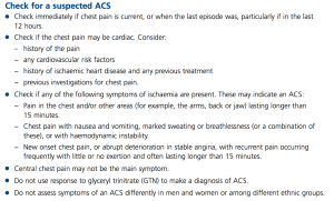
I was basing this on the criteria given on the NHS Clinical Knowledge Summary, and the correct answer was meant to be A, as the pain needs to last at least 15 minutes to be suspicious of ACS. In agreement with the NICE guidance, the GTN response is meaningless. Pain has to occur at rest or with minimal exertion (or with less and less exertion for crescendo angina) to be ACS.

NICE guidelines on ACS

I was about to put this in the app when I decided to cross check it with SIGN, purely out of interest. When considering the question of what signs or symptoms are useful in ACS:

“A high quality systematic review of 21 studies examined the usefulness of 16 different clinical signs and symptoms in the diagnosis of acute coronary syndromes. Taken in isolation, no single sign or symptom was discriminatory.”

OK, so perhaps by using a cluster of symptoms we can rationally justify saying “I think this patient has ACS”, even if there is no single dealmaker/breaker symptom. I read the paper that SIGN’s conclusion was based on to check this.

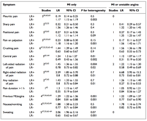
To a certain extent, you could. Let’s look at the likelihood ratios for the various symptoms in MI. The likelihood ratio is the factor you multiply the pretest odds by to get the posttest odds. If a patient had central, crushing pain lasting 1 hour, by virtue of this history his probability of MI has gone up by 1.24 x 1.44 x 1.3 = 2.32. It would have been more significant if the pain radiated to the right than the combination of those factors. I will emphasise that. Radiation of the pain to the right confers a LR of 2.59. This compares to 1.45 for radiation of pain to the left.

It becomes even more counter everything-we-have-been-taught when you look at the LR for MI or unstable angina. Radiation of the pain to the right has a LR of 6.68 (admittedly with a CI of 2.95 to 15.2), which dwarfs all the other factors in the history that we classically use. Nothing else has an average LR of even 2 or more.

MI symptoms and their usefulness

So it seems that perhaps we can use a combination of factors in the history, but we need quite a few of them. We should also respect right sided radiation as more worrying than left sided radiation.

(On a loosening of associations type trail of thought, RBBB is as common as LBBB in MI, and is associated with a worse in-hospital mortality. The SIGN guidance section 2.1 reflects this as well. It seems we’ve got our rights and lefts the wrong way round altogether in ACS.  Also, LBBB does not usually mean MI, even if you have no old ECGs to compare to.)

There were some symptoms that were helpful at reducing the LR. Pleuritic chest pain had a LR of 0.19, and pain on palpation gave 0.23. Examination findings were also useful. I used the mnemonic MR HOPE to remind me about what examination findings matter in MI: MR = mitral regurgitation, HO = hypotension (LR 3.06) and PE = pulmonary edema (LR 2.08)/heart failure signs.

Simple enough. Let’s suppose we now have two patients with central crushing chest pain lasting 1 hour. This has a LR of 2.32. If a patient had a massive pre-test probability before this i.e. lots of CV risk factors, then this is potentially more significant than in a patient with no risk factors at all. This is why the risk factors are so important for ? ACS. It is the risk factors that determine your post-history ACS probability as much as the history of the presenting complaint itself. That said, it would be a brave doctor to discharge either patient without a 12 hour troponin.

12 hour troponins are the bane of hospital bed managers around the country. You have well patients occupying an acute medical bed for the next 10 hours whilst the GI bleeder is seen in the corridor. I wonder if this approach could be used when it comes to cardiac enzymes on a low risk patient. Myoglobin rises within 2-4 hours. It is really nonspecific, but highly sensitive. This means that it could be useful for excluding the diagnosis if negative, but not much use if positive. It’s like d-dimers. In fact, just like d-dimers, if it comes positive, you need to do the “real test” (12 hour troponin). Think how many beds not having a pretty well person hanging around for a 12 hour troponin could save.

(I know emedicine does not like this approach. However, if we were to restrict ourselves to using it after at least 4 hours, and on low risk patients, I think it could work. Need to do more reading into it though.)

Conclusions:

1. Myoglobins to be the new d-dimers. You read it here first.

2. Right sided radiation to be respected more. And history always interpreted in light of the patient’s risk factors (or “pre history of presenting complaint probability”)

Anything intermittent in medicine is electrical

A lady in her late 50s with a background of metastatic colon cancer was referred by her GP with a Hb of 6.1 (MCV 89.8, WCC 20.9, Plt 117). This was noted because she had been complaining of worsening shortness of breath for the past week.

Her Hb the day before was 6.4, and her Hb 2 weeks ago was 11.6. She had had one previous transfusion when her Hb was restored from 5.3 to 9.4. Before and after that episode, she was generally around 10.5-12.

On arrival, she had a pulse of 130, a BP of 117/78 and was saturating 100% on air with a respiratory rate of 14. She had a temperature of 36.6.

I like to split the differential for shortness of breath according to the onset of symptoms. It would be very tempting to attribute the whole thing to anaemia, arrange a blood transfusion and leave it for the day team to follow up. I arranged for the transfusion to begin and then clerked her.

The shortness of breath was a gradual onset, and had been gradually progressing. Her shortness of breath had left her unable to walk more than 5m. This compares to a week ago, when she was able to walk around her supermarket with no problems.

She had intermittent chest pain, which was worse on inspiration. Shortness of breath, chest pain worse on inspiration, tachycardia and a PMH of metastatic cancer? I decided not to do any investigations into PE/DVT. This is either mad or brave. Here’s my thinking: The pain localised to her left lower ribs, directly over where the costochondral joints are. She was able to point to a specific point on her chest where the pain was. The pain lasted a maximum of 5 minutes, coming in waves when it was there. It occurred mostly in the morning on awakening. It was brought on by postural changes such as when getting out of bed and by overlying pressure. Between episodes, there was no pain at all. There was no cough, and no leg swelling/pain.

The other thing is that she was saturating at 100% on air, and still felt short of breath. This means that there is no problem getting oxygen from the atmosphere into the lungs, nor in getting oxygen from her lungs into her circulation. Of the causes of pleuritic chest pain, you would expect a pneumonia to affect the former, and a PE to affect the latter. The problem here was clearly quite different; there was not enough oxygen carrying capacity in her blood.

The shortness of breath was improved by lying flat, which counted against co-existing heart failure, which potentially could have been precipitated by anaemia. There were no infection pointers on history despite the raised WCC.

There was no history suggestive of GI, GU, PV, intracranial, intra-articular or other superficial bleeding.

She was generally quite an active lady for someone with metastatic cancer, and had no problems living with her husband in managing her ADLs. She was also a lifelong non smoker.

Her CVS, Resp and abdominal exam were all normal, except for the pulse of 130. Her mucous membranes were actually relatively moist, and she was talking comfortably in full sentences. There were no added noises in the chest, nor any swelling around the ankles/calves. There was no splenomegaly.

The question now became what caused the normocytic anaemia. Broadly, the causes can be split into a failure to make RBCs, or losing RBCs.

You fail to make RBCs either because the marrow is rubbish or because the marrow is not stimulated properly.

You lose RBCs in bleeding, hypersplenism and haemolysis. Haemolysis splits into congential and acquired. Acquired haemolysis splits into autoimmune and mechanical. I will make a diagram for all this at some stage.

I was struggling to come up with an adequate explanation for her acute episode of pretty severe normocytic anaemia. I decided the best plan would be to treat her anaemia, and then review what symptoms if any she had post transfusion. I went to see the consultant ready to defend the decision to avoid D-dimers or a chest xray in this case unless she does not respond to the transfusion.

I was relieved and heartened by the consultant agreeing with this. So many AMU clerkings get ‘routine’ chest xrays, and I do not understand this. If you are struggling to put something meaningful on the chest xray request card, surely that should be an indication to think twice about whether or not it is justified, not to make up that you heard crackles on the left base on examination as I have seen and been encouraged to do by several doctors.

As for the cause of the anaemia, the consultant asked me to link cancers in general to normocytic anaemia, going back to pathology. Having just tutored in the 6 hallmarks of cancer, I thought of tissue invasion and the bleeding risk. This was not was he was after. He asked me to think again about the metastatic nature of the cancer. It went to the bone, and then my eureka moment. The bone marrow. Any cancer causing lytic lesions in the bone could eventually invade the bone marrow itself.

As for the intermittent symptoms, my consultant told me he was not worried about PE or pneumonia.

“Only four things cause intermittent symptoms in medicine, and they are all electrical.”

“Electrical?” 

“Yes, electrical. Seizures are electrical. Neurological pain is electrical. Arrythmias are electrical. Muscle spasms are electrical. Pretty much all intermittent symptoms are one of these four. ”

“What about colic?”

“That’s surgical.”

The more I thought about it, the more I agreed. There were some exceptions, like osteoarthritis pain being worse on exertion and then clearing, or calf claudication worse on exertion. I think this concept could be improved by adding the word ‘spontaneous’. This would exclude symptoms that clearly arise following a provoking factor. I am struggling to come up with an exception to the rule that all spontaneously intermittent symptoms in medicine are electrical in nature, and are one of a seizure, neurological pain, an arrhythmia or a muscle spasm.

Any ideas?

Approach to a patient with a 7.6cm AAA

Things were livened around 11am up by a call from the ultrasonographer. A man in his late 60s year had undergone an abdominal ultrasound at the request of the consultant haematologist, with a view to confirming his impression of splenomegaly. The ultrasonographer instead found a 7.6cm aneurysm, and wanted me to make a plan.

I decided to go and see the patient in the room, and asked the nurses to get some obs on him. He was symptom free, comfortable at rest and his obs were stable. He had back pain for the past 12 months, which was relieved by leaning forward. There was no leg pain. This seemed consistent with spinal stenosis. There had been no recent progression.

His PMH showed COPD as well as RAEB (refractory anaemia with excessive blasts), with a generally poor exercise tolerance of a few metres. Poor exercise tolerance, low platelets, chronic anaemia…surely not the best candidate for surgery? Where are the guidelines for this scenario?

It turns out there is a lack of data in this field. “Guidelines for the treatment of abdominal aortic aneurysms: Report of a subcommittee of the Joint Council of the American Association for Vascular Surgery and Society for Vascular Surgery”, notes this, and offers the following:

Decision making involved in selecting patients for AAA repair is influenced primarily by estimates of (1) aneurysm rupture risk, (2) elective operative mortality risk, (3) life expectancy, and (4) patient preference. In the absence of truly accurate data regarding many of these variables, decision making is often a complex and uncertain process. It is increasingly recognized that patient preference, after a complete review of options and anticipated results (true informed consent), must be a very important component in this decision-making process.

What is the rupture risk? The same paper offers the following. Bear in mind the normal abdominal aorta has a diameter of 2cm, and 3cm and above defines an aneurysm.

AAA diameter (cm) Rupture risk (%/y)
<4 0
4-5 0.5-5
5-6 3-15
6-7 10-20
7-8 20-40
>8 30-50

Interestingly, although AAAs are twice as common in men than in women, women are at 4 times the risk of rupture compared to men, and a greater proportion of female ruptures are fatal. The 5.5cm diameter cut off for elective surgery in the UK was originally designed for men, and this number should perhaps be lower in women.

What were my patient’s risk factors for AAA? I had always been taught that it was basically the atheroscelerotic risk factors, with an emphasis on smoking and hypertension. Then there were the medical school favourites like Ehlors Danlos and Marfan’s. I have found out today that diabetes is protective against AAA, and that there is no association with cholesterol levels. This suggests that the disease process is not simply atherosclerosis. In addition, COPD is an independent risk factor from smoking.

I thought back to the non rupture complications of AAA, and remembered about the microemboli that can be thrown off. Our patient had perfectly good pulses bilaterally, and no evidence of emboli in the feet.

At this stage, I was stuck with an anxious man who has just been told he has a large blood vessel in his abdomen that could burst at any second as well as two medical students and a nurse looking at me expectantly. My belief in guardian angels was restored when my bleep went off. I was delighted to hear the voice of my SHO, who had thought to bleep me just to see what’s up. The vascular reg came down to discuss it with the patient, and an urgent outpatient appointment was arranged to go through his options more fully once he had more time to reflect.