Left and right heart failure do not exist

Last Thursday, the cardiology consultant argued that dividing heart failure symptoms into left sided and right sided did not make sense. After all, if the left pump is failing, there will be backwards and forwards failure. The forwards failure will reduce venous return, which will reduce the right ventricular output by reducing preload. The backwards failure will increase the pulmonary pressure, and will eventually reduce the right ventricular output by increasing afterload. It is therefore impossible to have purely isolated symptoms of univentricular heart failure, and making the distinction between left and right sided failure clinically is meaningless.

This was shocking to me and the students. We had been taught so much about left sided vs right sided failure. I decided to go back to basic science and evidence to see if I could work it out for myself.

What is the role of the right ventricle?

In 1943 and seemingly oblivious to some sort of war going on, Starr et al ablated the RV in open chest dogs with “a red hot soldering iron,” and found little alteration in systemic circulation or venous pressure.

Starr I, Jeffers WA, Meade RH. The absence of conspicuous increments of venous pressure after severe damage to the RV of the dog, with discussion of the relation between clinical congestive heart failure and heart disease. Am Heart J. 1943;26:291-301.  

At the highly misogynist time, everyone thought that this meant the right ventricle was little more than a pretty bride to the hunky husband of the left ventricle, which was the dominant pump determining cardiac output.

And so it remained, until further studies showed that this may be true in normal health, but the moment pulmonary pressures increase the performance of the right ventricle falls off. This situation arises in cor pulmonale, acute PE (which can be thought of as acute cor pulmonale)…and significant left ventricular failure.

Brooks H, Kirk ES, Vokonas PS, Urschel CW, Sonnenblick EH. Performance of the right ventricle under stress: relation to right coronary flow. J Clin Invest. 1971;50:2176-83

More recently, it was shown that having a right ventricular infarct on top of your (usually inferior) left sided infarct was associated with a worsened mortality and morbidity.

Zehender M, Kasper W, Kauder E, Schonthaler M, Geibel A, Olschewski M, et al. Right ventricular infarction as an independent predictor of prognosis after acute inferior myocardial infarction. N Engl J Med. 1993;328:981-8. 

Interestingly, they found that having a right ventricular infarct on top of your LV infarct led to worsened hypotension, which is a classical ‘left sided’ symptom from forwards failure. This study noted this effect was independent of the size of the left sided infarct. The findings have been replicated where shock was more common in those patients who had right sided failure.  So, the right ventricle’s function is important, especially when the left ventricle is impaired. This is consistent with our consultant’s message at the start that the failure of one ventricle invariably messes up the other.

LV function is also a direct determinant of RV ejection, via the effects of LV function on the contraction of the interventricular septum and the wringing action of the LV.

In the same article, we can see how impaired RV function can lead to a dilated and hypertrophied right ventricle, which in turn can shift the septum towards the left and impair LV filling.

A match made in the pericardium

RV hypertrophy occurs as a response to increased wall stress. The growing RV forces the IV septum into the left ventricle, as the pericardium prevents the outwards growth of the right ventricle.

Does this answer the question of whether or not the forwards/backwards failure means you always have the same symptoms anyway?

Although we can see that failure of one ventricle leads to failure of the other, this is not the same as saying there is no clinical distinction between the two. I’m still to be convinced that there is no role at all for distinguishing the two types of failure clinically. I struggled to find any sources which suggest there is no value in distinguishing the two clinically, and that both must invariably exist together. That could be because it’s hard to search for a negative i.e. pages which do not state left and right sided symptoms separately. I still feel that isolated left/right ventricular failure is a real thing (which may progress to biventricular failure) but will keep open minded about this and be ready to claim I blogged about it first if and when the death of left/right failure as a classification becomes mainstream.
Getting tied up with the spinal cord

Spinal cord compression

Thanks to firm 3 for the enthusiasm at today’s bedside teaching.

We saw a gentleman in his 90s who presented with difficulty walking for the past 3 days. He also noted numbness in his right leg distal to the shin over this time. There was no pain, subjective complaints of weakness or change to his bowel/bladder function. He had a recent diagnosis of lung cancer and the concern was about a possible compression of the spinal cord.

We went through the ways a metastasis to the spine may present. We split the symptoms into those related to the bony invasion and those related to the compression of neural bits.

Bony invasion

Severe, horrendous back pain that keeps the patient up at night

Thoracic/cervical pain

Point tenderness

Pain worsened by direct pressure

Pain worsened by straining

Neurological involvement

Motor loss

Sensory loss

Autonomic loss, which can present as bowel/bladder disturbance (often ileus or retention)

‘Off legs’, non specific difficulty walking

Super red flags for cauda equina

Saddle anaesthesia – but be careful about how you ask this question. There is a lesson to be learnt from the case report in the BMJ where a GP simply asked if the patient was numb, which led to the diagnosis being delayed. I have also blogged about the case I discussed with you about a possible cauda equina during my psychiatry job. Ask if it feels different when you sit down or wipe yourself with the toilet paper, not simply whether or not the area is numb. Perianal sensory disturbance occurs in stage 2 of cauda equina, which is much more amenable to treatment than stage 4, where there is perianal sensory loss.

Back pain – one of the earliest signs, but non specific.

Bilateral or alternating leg pain – bilateral or alternating leg pain should always make you think spinal cord or cauda equina. Radiculopathy from simple disc herniation is nearly always unilateral.

Note that we are not saying that all disc hernations are benign. You should advise a patient with disc herniation that although the majority self resolve with no problems, he or she must seek immediate medical attention if symptoms of cauda equina occur. The most common cause of cauda equina is actually disc herniation (occurs in 2% of all disc herniations).

Motor/sensory loss – depending on the exact nerve roots affected. In cauda equina there will only be LMN signs; in conus medullaris there may be both UMN and LMN signs.

Bowel symptoms – any change in bowel habits, but especially ileus symptoms i.e. not passing motions. On PR examination there may be loss of anal tone.  This is a very late sign.

Bladder symptoms – usually retention, and this is a late sign.

What do you do if you think a patient has a spinal metastasis?

1. Assess the severity. The three grades of severity correspond to a) just bony pain b) neurological involvement and c) if emergency treatment could be planned for this condition e.g. cauda equina

2. Discuss with your senior immediately. This patient may need high dose dexamethasone to reduce the swelling around the tumour. There is a limited role for bisphosphonates in breast cancer and myeloma (and sometimes prostate cancer).

3. Ensure adequate analgesia

4. Arrange MRI with the appropriate degree of urgency having discussed with your senior. They may consider issues like the patient’s desired ceiling of care, life expectancy and fitness for intervention.

According to NICE:

Perform MRI of the whole spine in patients with suspected MSCC, unless there is a specific contraindication. This should be done in time to allow definitive treatment to be planned:

– within 1 week of the suspected diagnosis in the case of spinal pain suggestive of spinal metastases,

– within 24 hours in the case of spinal pain suggestive of spinal metastases and neurological symptoms or signs suggestive of MSCC

– occasionally sooner if there is a pressing clinical need for emergency surgery

The treatment from here is essentially surgical, radiotherapy or conservative.


Cord lesions

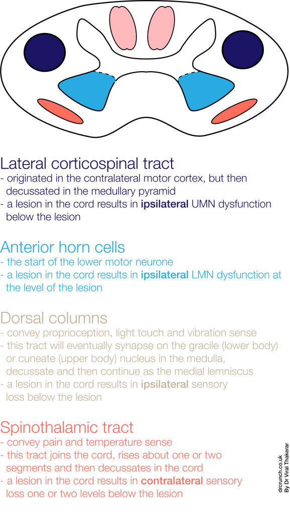
I thought it would be helpful to recap the anatomy and work out the clinical consequences of some of the cord syndromes we spoke about today:

Spinal cord anatomy broken down into just the clinically relevant bits


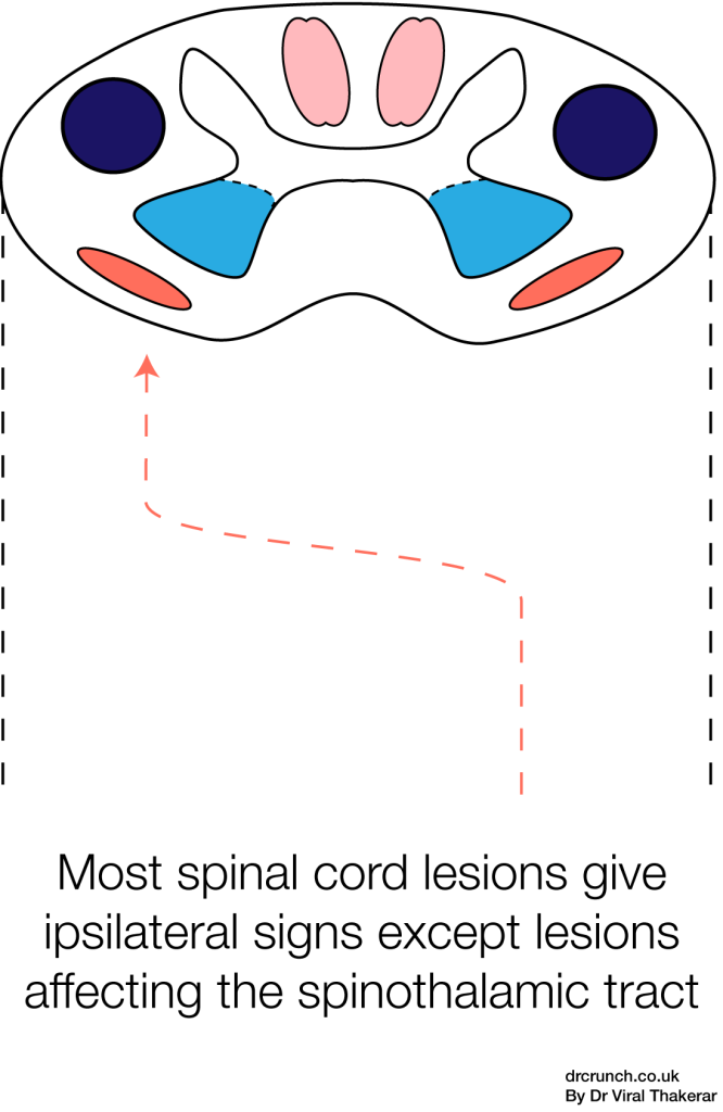
The spinothalamic tract is the odd one out.

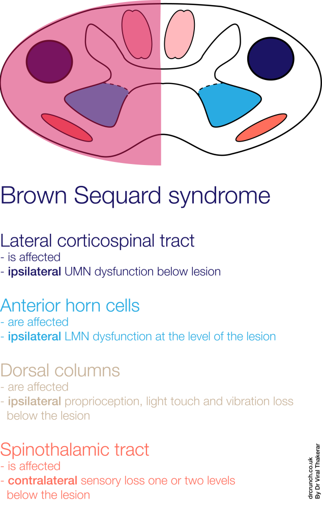
The Brown Sequard syndrome
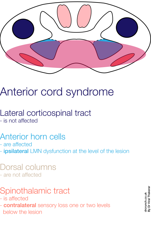
Anterior Cord Syndrome simplified.