GCS: Causing more harm than good since 1974

Triage notes: 61 yo man, reducing GCS. PMH myelofibrosis

Reduced GCS? Uh oh. What could be going on in that tortured skull?

Is the first thing I would normally think with a reduced GCS. I have learnt to use these piecemeal snippets of information to guide rather than dictate the direction of my history taking. What exactly do they mean by a reduced GCS?

From the ambulance clerking:

“Eyes not open except to voice. Obeying commands. Orientated and talking.”

If you tried to GCS me at 3am I’d probably be the same. Was this patient actually having a neurological problem, or was he just really tired?

His background of myelofibrosis made me suspicious for an acute on chronic anaemia, as well as infection from immunosuppression. The A&E ‘in-and-out’ part of me wanted to just CT Head him because it seems rude to not to on a patient with an apparently deteriorating GCS. The ‘history first, examination second, investigations third’ part of me decided to just hold back a second and be sure that there is a real indication for CT Head.

I decided to take a detailed history first. I learnt that this man had been down the pub just 4 days ago and enjoyed a meal there, socialising with his family and friends. Over the next few days, he spent more time in the chair and not moving around so much. He got short of breath on progressively less and less exercise, to the point where he was taking his time up the stairs. The night before the day of admission he went to sleep at 9pm. The next morning, he was unrousable according to his wife.

Over this time, he had intermittent fevers and night sweats, but had had these for ‘years’. He had flu-like symptoms a few weeks earlier but these had resolved a week before. There was no leg swelling or shortness of breath on lying flat.

There was no focal weakness or sensory loss, or changes to his speech. However, he had become weak all over according to his wife. He had no headache, vomiting or changes to his vision.

On examination, he seems dehydrated, with a pulse of 85. He had no focal neurological signs and his pupils were equal and reactive. His eyes were closed, but opened on command. I felt he was not actually unresponsive and was instead just keeping his eyes shut unless needed. He was a little weak with all movements.

I took bloods for possible sepsis and a VBG, which showed he was not anaemic and had a lactate of 1.4 mmol/L and an Hb of 140 g/L, which ruled out anaemia. I decided to give him some fluids and broad spectrum IV antibiotics whilst awaiting the FBC result, and if he did not improve on this to consider an intracranial cause. I also sent off TFTs.

One hour later, as I saw him wheeled out the corridor to the medical admission ward, he was a different man altogether. He was waving and talking. The wife said that she got her husband back.

The GCS revisited

This experience made me think about how the GCS maybe gets abused in the healthcare system. Teasdale and Jennet invented the GCS in 1974 as a tool for monitoring the coma level in patients on a neurosurgical ward. They tried in vain in 1978 to empahsise that ‘we have never recommended using the GCS alone, either as a means of monitoring coma, or to assess the severity of brain damage or predict outcome.’

Even more bizarre is how we add up the components. Is a patient who has moved from being unable to localise pain to having a flexion withdrawal response as much of a deterioration as a patient who was talking to you but is now disorientated? The individual parts just aren’t comparable to each other, as the original authors wrote. The components of the scale were simply ranked e.g. localises to pain is one place worse than obeying commands. This is completely different to giving them scores, which implies equal spacing between them.

In addition, to add up the components implies that the scale should be weighted 6/15 to motor, 5/15 to vocal and 4/15 to eyes. This does an injustice…to motor. The motor part is the most important part of the scale, and similar validity for the usual applications of the GCS can be found using a simplified motor only version of the GCS.

You can read an excellent article by Dr Green about the problems with how we use GCS. One bit I liked from this article was where they looked at how meaningless the summarative GCS could be for prognosis:

The 13 possible GCS values can include 120 combinations of its components. A GCS score of 4 predicts a mortality rate of 48% if calculated 1,1,2 for eye, verbal, and motor, a mortality of 27% if calculated 1,2,1, but a mortality of only 19% if calculated 2,1,1.

So what should we use instead?

How about the Simplified Motor Scale, also known as TROLL? Given neurosurgeons get GCS wrong nearly half the time (Bassi S, Buxton N, Punt JA, et al. Glasgow Coma Scale: a help or a hindrance? Br J Neurosurg. 1999;13:526-539), surely it is safer to use a tool which has less margin for subjectivity and as much validity for head injuries at least?

Had we used the SMS, this patient would have been admitted as fatigued/dehydrated rather than reduced GCS, which would have been far more appropriate.
Syncope query cause? Risk stratification is the aim of the game

As an FY1 when I saw syncope I saw my job as taking a nice pre, during and post history of the event and working out what the most likely cause was. This still mostly holds, but I realise the job of the A&E doctor is both less than this and yet beyond this.

I recently saw a man in his 60s present as ? head injury. He had just left the shower and felt like he was about to vomit. He then sat down on the toilet (not using the toilet but just sitting on it) and a few seconds later fainted. He was unconscious for about 20 seconds as witnessed by a family member. There was no incontinence, tongue biting or unusual movements. There was no chest pain or palpitations before the fall, and he felt fine afterwards. He may have banged his head on the radiator on the way down, but was otherwise fine and complained of no injuries.

He was being investigated for obstructive jaundice at the time.

His past medical history included Type II diabetes, a meningioma excised 5 years ago and purportedly hypertension. His medications were metformin, ramipril, simvastatin and loratidine.

Neurologically he was intact apart from a right pupil of 4mm compared to a left pupil of 3mm. There was a scar from a previous resection of a meningioma.

Cardiovascular examination was unremarkable. Postural blood pressures showed a significant drop (133/76 versus 101/74).

An ECG showed sinus bradycardia at 54.

FBC, U&Es and CRP were all unremarkable. LFTs confirmed obstructive jaundice.

Using the four groups of 3 trick, I worked through the list.

Cardiac: Arrythmia, ACS, outflow obstruction (basically aortic stenosis or HOCM)

Neurological: Seizure, stroke/TIA, raised ICP causes (especially bleeds in the acute setting)

Neurologically mediated: Vasovagal, carotid sinus hypersensitivity, cough/micturition syncope

Oddballs: (I think of these as ‘PH, P, H’): Postural Hypotension, PE, Hypoglycemia

I felt that this patient probably had a syncope related to his postural hypotension from ramipril or autonomic failure from diabtes, with an arrhythmia less likely.

The A&E registrar decided that the patient should go under the medics.

When I made the referral, I expected to be quizzed as to my likely cause for the syncope. Instead, I got asked quite simply: “What’s his risk?”

His risk of what? Of each of the causes of syncope?

“Risk…?” I said

“His risk.”

I didn’t realise what she was talking about. Was this specifically for arrhythmia?

“Well he is in sinus at 54. The ECG was otherwise unremarkable.”

“Has he got any cardiac failure?”

Ah, getting warmer. This is the risk for arrhythmia/cardiac causes.

“Not really, though he is a diabetic with hypertension so has a decent ischaemic risk.”

“Fine”  she said in a voice that was anything but fine with it.

I decided to find out what this risk was. It turns out there are scoring systems for predicted the risk of a cardiac cause for the syncope in the ED. You can use the ROSE score  but even this isn’t fully accepted. It places emphasis on the BNP, which is quite expensive and not often measured in the ED.

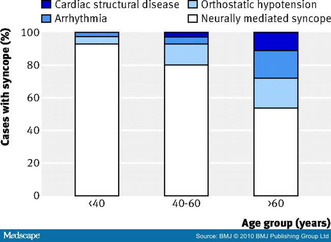
Another approach to picking out the patients at high risk of cardiac syncope is to use the patient’s age to mentally risk stratify them:

pre test syncope causes

You can see how past the age of 60 non simple causes become a lot more prevalent. In fact, the cardiac causes of syncope have a bimodal distribution; the smaller peak is young people with inherited cardiac conditions and the larger peak is people over 65.

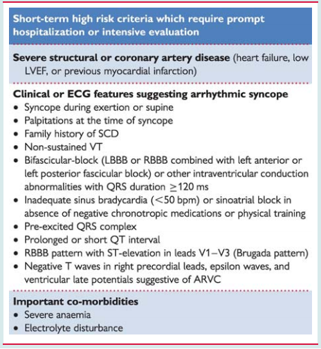
You can then use the ESC guidelines to get an idea of the risk for a cardiac cause. This could then inform the medical team about the likelihood that there is a cardiac cause worthy of further investigation.

risk

It’s a change from how I used to think about things a year ago, where I was focused on making the diagnosis as accurately as possible. Now I realise the job is to work out where the patient is heading next, even if we cannot quite label the condition at the moment.

By Viral Thakerar

How to distinguish a true Babinski from tickly feet

Switch the sound off and watch the clip above. Is this a true positive Babinksi, or simply a withdrawal response? How did you tell? And could you explain it to someone else?

But everyone has tickly feet…

If you were to stroke the soles of most people’s feet with a blunt object, they wouldn’t like it. Most people will make some movement to get away from the annoying stimulus. The question I posed to the medical students was how do you distinguish a desire to move the foot away from the stimulus from a true positive Babinksi.

The answer comes from reading Joseph Babinski’s original description of the sign. When the lateral aspect of the foot was stroked (not scraped or anything painful) he observed:

In normal people, there was flexion synergy of the thigh, knee and toes

BUT

In those with hemi or monoplegia of central origin there was flexion of the thigh and knee but upwards extension of the toes.

Why so?

Let’s go back to being a newborn baby. If there’s something annoying on your foot, you want to shorten your limb to get away from it. The spinal cord has this built in functionality out of the box, and has a reflex to deal with this situation without any input from the pyramidal tract. This involves flexing everything in the lower limb except your toes, which you extend. This is called flexion synergy.

Why is it called flexion synergy when the toes are clearly not flexing? The toes are clearly not in synergy with everything else.

True. When you think things through in limb anatomy, the naming conventions make no sense (e.g. naming movement of the thumb). What we call extending the toes makes the limb shorter, which is unusual for extension. The built in spinal cord level response is a flexion withdrawal and this involves extending the toes to shorten the limb. Maybe it was all flexion once upon an ancient evolutionary path when our toes pointed in a different direction.

So what happens as we age?

Around the time the infant starts walking, the pyramidal tract matures enough to suppress this flexion synergy response. The contraction of the tensor fascia lata of the thigh and the knee may remain, but the toes no longer respond as part of the flexion synergy.

The toes don’t respond? I thought they were down going normally?

They don’t respond as part of the flexion synergy. The reason they go downwards is to do with a local cutaneous reaction. That is why if you elicit the Babinski using an alternative method e.g. Oppenheimer’s sign, you don’t get the toes curling down but you would get the other components of the sign.

Hold on, Oppenheimer?

There are over 30 ways of eliciting the Babinksi. I have no intention of learning them all, but the Oppenheimer is a useful alternative to have in your arsenal as it doesn’t elicit a tickly response so can be helpful for sensitive soles.

I see what you did there with the last two words of that paragraph.

Thank you.

So what features help distinguish a true positive Babinski?

1. Upward movement of the toe is pathological only if caused by contraction of extensor hallicus longus muscle. Beware of labelling upwards movements of the toes due to ankle flexion as a Babinski.

2. Contraction of extensor hallicus longus muscle is pathological only if it occurs synchronously with reflex activity in other flexor muscles. The easiest one to observe is the tensor fascia lata of the thigh – see you the YouTube video.

3. A true up-going toe sign is reproducible, unlike voluntary withdrawal. I was often taught as a medical student that you can ‘tire out’ a Babinski. In reality, you are much more likely to tire out a voluntary withdrawal than a Babinski.

Go back to the video at the top that demonstrates a very clear true Babinski. See if you can identify features one and two. Note how the flexion synergy occurs at the same time as the great toe extension.

Further reading:  An excellent review by Van Gijn