Know your rights when it comes to ACS

You know that feeling on your first day of A-Level chemistry when you find out everything they told you in GCSE was a lie? I felt something similar when I tried to find out which symptoms in ACS are actually helpful for ruling in/out the disease. It seems that few of the symptoms we are classically taught about are that useful, and the few that are reasonably good are not the ones we are taught about.

I first realised this whilst writing a new SBA for my app. I wanted to highlight what the criteria for suspecting ACS really were. This was based on an experience about a month ago where for about a week I was asked to see multiple patients/referrals from GPs for patients with chest pain that sounds like ‘classic’ ACS…until you clarify that the pain lasted seconds.

The SBA went like this:

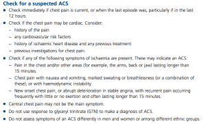
A 53 year old man comes in with chest pain. When you assess him, the pain has subsided and he feels well. It was a central pain, occurring at rest and lasting about 5 minutes. There was radiation to the right, and there was no response to GTN. Which of these features makes the diagnosis of ACS most unlikely?
A Duration of 5 minutes
B Lack of response to GTN
C Spontaneous resolution of pain combined with feeling well
D Pain occurred at rest
E Radiation to the right

I was basing this on the criteria given on the NHS Clinical Knowledge Summary, and the correct answer was meant to be A, as the pain needs to last at least 15 minutes to be suspicious of ACS. In agreement with the NICE guidance, the GTN response is meaningless. Pain has to occur at rest or with minimal exertion (or with less and less exertion for crescendo angina) to be ACS.

NICE guidelines on ACS

I was about to put this in the app when I decided to cross check it with SIGN, purely out of interest. When considering the question of what signs or symptoms are useful in ACS:

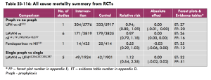
“A high quality systematic review of 21 studies examined the usefulness of 16 different clinical signs and symptoms in the diagnosis of acute coronary syndromes. Taken in isolation, no single sign or symptom was discriminatory.”

OK, so perhaps by using a cluster of symptoms we can rationally justify saying “I think this patient has ACS”, even if there is no single dealmaker/breaker symptom. I read the paper that SIGN’s conclusion was based on to check this.

To a certain extent, you could. Let’s look at the likelihood ratios for the various symptoms in MI. The likelihood ratio is the factor you multiply the pretest odds by to get the posttest odds. If a patient had central, crushing pain lasting 1 hour, by virtue of this history his probability of MI has gone up by 1.24 x 1.44 x 1.3 = 2.32. It would have been more significant if the pain radiated to the right than the combination of those factors. I will emphasise that. Radiation of the pain to the right confers a LR of 2.59. This compares to 1.45 for radiation of pain to the left.

It becomes even more counter everything-we-have-been-taught when you look at the LR for MI or unstable angina. Radiation of the pain to the right has a LR of 6.68 (admittedly with a CI of 2.95 to 15.2), which dwarfs all the other factors in the history that we classically use. Nothing else has an average LR of even 2 or more.

MI symptoms and their usefulness

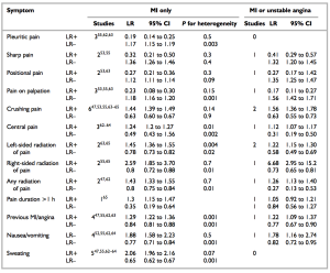
So it seems that perhaps we can use a combination of factors in the history, but we need quite a few of them. We should also respect right sided radiation as more worrying than left sided radiation.

(On a loosening of associations type trail of thought, RBBB is as common as LBBB in MI, and is associated with a worse in-hospital mortality. The SIGN guidance section 2.1 reflects this as well. It seems we’ve got our rights and lefts the wrong way round altogether in ACS.  Also, LBBB does not usually mean MI, even if you have no old ECGs to compare to.)

There were some symptoms that were helpful at reducing the LR. Pleuritic chest pain had a LR of 0.19, and pain on palpation gave 0.23. Examination findings were also useful. I used the mnemonic MR HOPE to remind me about what examination findings matter in MI: MR = mitral regurgitation, HO = hypotension (LR 3.06) and PE = pulmonary edema (LR 2.08)/heart failure signs.

Simple enough. Let’s suppose we now have two patients with central crushing chest pain lasting 1 hour. This has a LR of 2.32. If a patient had a massive pre-test probability before this i.e. lots of CV risk factors, then this is potentially more significant than in a patient with no risk factors at all. This is why the risk factors are so important for ? ACS. It is the risk factors that determine your post-history ACS probability as much as the history of the presenting complaint itself. That said, it would be a brave doctor to discharge either patient without a 12 hour troponin.

12 hour troponins are the bane of hospital bed managers around the country. You have well patients occupying an acute medical bed for the next 10 hours whilst the GI bleeder is seen in the corridor. I wonder if this approach could be used when it comes to cardiac enzymes on a low risk patient. Myoglobin rises within 2-4 hours. It is really nonspecific, but highly sensitive. This means that it could be useful for excluding the diagnosis if negative, but not much use if positive. It’s like d-dimers. In fact, just like d-dimers, if it comes positive, you need to do the “real test” (12 hour troponin). Think how many beds not having a pretty well person hanging around for a 12 hour troponin could save.

(I know emedicine does not like this approach. However, if we were to restrict ourselves to using it after at least 4 hours, and on low risk patients, I think it could work. Need to do more reading into it though.)

Conclusions:

1. Myoglobins to be the new d-dimers. You read it here first.

2. Right sided radiation to be respected more. And history always interpreted in light of the patient’s risk factors (or “pre history of presenting complaint probability”)

Venous thromboembolism: maintaining the risk

One of the banes of modern NHS hospital life is filling in VTE assessments. These have become ever more intrusive since June 2010 as a result of the Department of Health decreeing that hospitals will be fined unless 90% of all patients have a VTE assessment done. Is this evidence based stick wielding, or simply political point scoring to claim that the DoH is being proactive about a potentially preventable cause of death?

This is the letter that first got me thinking. There is no doubt about the benefits of thromboprophylaxis in surgical patients, but the evidence for medical admissions is less clear. It seems that for every 400 medical patients treated with LMWH, one symptomatic PE is prevented at the cost of two serious haemorrhages.

We also only give prophylactic LMWH whilst medical patients are in hospital, yet DVT/PEs can form up to 6 weeks after an acute inflammatory condition.

This looks bad. Let’s see if there’s anything else to fly the flag for VTE prophlyaxis.

It turns out there is a greater success rate against DVTs – from the same review, for every 16 medical patients given thromboprophylaxis, one DVT is prevented.

We know that there is a 6% mortality rate within one month of diagnosis of a DVT, and a 12% mortality rate within one month of a PE, assuming adequate anticoagulation. Interperating this is complicated by the fact that having a DVT/PE is a pretty strong marker of generally being unwell when you think of the risk factors: cancers (especially metastatic), advanced age, worsening mobility etc. This makes it hard to be certain how much the DVT/PE actually contributed to the death compared to how much the DVT/PE was a marker of impending death from co-morbidities. The paper generally tried to use autopsy proved PE as a cause of death, which should help reduce other causes of death being ascribed to PE. It does not seem to include the mortality from hemorrhage from the anticoagulation, but I could not be certain from reading it.

For every 400 medical patients given LMWH, at one month we have saved 6% of the 25 who did not get a DVT thanks to us, and about 12% of 1 patient who was spared a PE. Total number of patients saved = 0.06 x 25 + 0.12 x 1 = 1.62 patients

The 26 people who just got a diagnosis of DVT/PE would have been on full dose anticoagulation for that month, at a monthly bleeding risk of 0.3% for a major bleed and just over 0.06% for a fatal bleed. Over three months, this is nearly 1% of the patients having a major bleed and 0.2% having a fatal bleed.

(3 months’ anticoagulation is what most proximal DVTs get. I know I only could find the mortality figures for 1 month and I’m using 3 months’ anticoagulation. I can’t find the perfect papers for this. Sincere apologies.)

Total of number of patients saved from having a major bleed by preventing the DVT/PE = 0.01 x 26 = 0.26, and 0.002 x 26 = 0.05 patients saved from a fatal bleed.

The risk of LMWH is bleeding, of which two per 400 were major. It’s not very clear what is defined as major in the summary.

In summary of this completely ad hoc and massively flawed analysis, out of 400 patients given LMWH, we probably saved 1.6 patients or so from a clot based death. We probably caused about 2 to have a major bleed, but saved 0.26 major bleeds in the process by sparing them anticoagulation to treat a clot.

NICE themselves did a better job of it, and even they concluded that in medical patients, there was no overall benefit at all from VTE prophylaxis. I can’t tell if the studies they used considered the effects of sparing patients from full dose anticoagulation for months on end if you prevent a clot. This is on page 360 of the full guidance:

NICE: Maintaining the evidence

That said, the response in BMJ letters made me realise I may have been a bit too hasty to completely disregard the other side to the debate.

What do you think?

(To add  an off topic point that does not follow from anything that I have just written, recent immobility that is worse than the patient’s normal state is a significant risk factor for VTE. That said, being generally immobile is not significant, especially if they have been that way for over 4 months. This is something I had not appreciated before.

More mud in the water. I’m glad I’m on surgery now so I can just fill VTE assessments without thinking about how we may be pawns being manipulated by politicians eager to prove that they are doing something about something important somewhere without any evidence base.)

Anything intermittent in medicine is electrical

A lady in her late 50s with a background of metastatic colon cancer was referred by her GP with a Hb of 6.1 (MCV 89.8, WCC 20.9, Plt 117). This was noted because she had been complaining of worsening shortness of breath for the past week.

Her Hb the day before was 6.4, and her Hb 2 weeks ago was 11.6. She had had one previous transfusion when her Hb was restored from 5.3 to 9.4. Before and after that episode, she was generally around 10.5-12.

On arrival, she had a pulse of 130, a BP of 117/78 and was saturating 100% on air with a respiratory rate of 14. She had a temperature of 36.6.

I like to split the differential for shortness of breath according to the onset of symptoms. It would be very tempting to attribute the whole thing to anaemia, arrange a blood transfusion and leave it for the day team to follow up. I arranged for the transfusion to begin and then clerked her.

The shortness of breath was a gradual onset, and had been gradually progressing. Her shortness of breath had left her unable to walk more than 5m. This compares to a week ago, when she was able to walk around her supermarket with no problems.

She had intermittent chest pain, which was worse on inspiration. Shortness of breath, chest pain worse on inspiration, tachycardia and a PMH of metastatic cancer? I decided not to do any investigations into PE/DVT. This is either mad or brave. Here’s my thinking: The pain localised to her left lower ribs, directly over where the costochondral joints are. She was able to point to a specific point on her chest where the pain was. The pain lasted a maximum of 5 minutes, coming in waves when it was there. It occurred mostly in the morning on awakening. It was brought on by postural changes such as when getting out of bed and by overlying pressure. Between episodes, there was no pain at all. There was no cough, and no leg swelling/pain.

The other thing is that she was saturating at 100% on air, and still felt short of breath. This means that there is no problem getting oxygen from the atmosphere into the lungs, nor in getting oxygen from her lungs into her circulation. Of the causes of pleuritic chest pain, you would expect a pneumonia to affect the former, and a PE to affect the latter. The problem here was clearly quite different; there was not enough oxygen carrying capacity in her blood.

The shortness of breath was improved by lying flat, which counted against co-existing heart failure, which potentially could have been precipitated by anaemia. There were no infection pointers on history despite the raised WCC.

There was no history suggestive of GI, GU, PV, intracranial, intra-articular or other superficial bleeding.

She was generally quite an active lady for someone with metastatic cancer, and had no problems living with her husband in managing her ADLs. She was also a lifelong non smoker.

Her CVS, Resp and abdominal exam were all normal, except for the pulse of 130. Her mucous membranes were actually relatively moist, and she was talking comfortably in full sentences. There were no added noises in the chest, nor any swelling around the ankles/calves. There was no splenomegaly.

The question now became what caused the normocytic anaemia. Broadly, the causes can be split into a failure to make RBCs, or losing RBCs.

You fail to make RBCs either because the marrow is rubbish or because the marrow is not stimulated properly.

You lose RBCs in bleeding, hypersplenism and haemolysis. Haemolysis splits into congential and acquired. Acquired haemolysis splits into autoimmune and mechanical. I will make a diagram for all this at some stage.

I was struggling to come up with an adequate explanation for her acute episode of pretty severe normocytic anaemia. I decided the best plan would be to treat her anaemia, and then review what symptoms if any she had post transfusion. I went to see the consultant ready to defend the decision to avoid D-dimers or a chest xray in this case unless she does not respond to the transfusion.

I was relieved and heartened by the consultant agreeing with this. So many AMU clerkings get ‘routine’ chest xrays, and I do not understand this. If you are struggling to put something meaningful on the chest xray request card, surely that should be an indication to think twice about whether or not it is justified, not to make up that you heard crackles on the left base on examination as I have seen and been encouraged to do by several doctors.

As for the cause of the anaemia, the consultant asked me to link cancers in general to normocytic anaemia, going back to pathology. Having just tutored in the 6 hallmarks of cancer, I thought of tissue invasion and the bleeding risk. This was not was he was after. He asked me to think again about the metastatic nature of the cancer. It went to the bone, and then my eureka moment. The bone marrow. Any cancer causing lytic lesions in the bone could eventually invade the bone marrow itself.

As for the intermittent symptoms, my consultant told me he was not worried about PE or pneumonia.

“Only four things cause intermittent symptoms in medicine, and they are all electrical.”

“Electrical?” 

“Yes, electrical. Seizures are electrical. Neurological pain is electrical. Arrythmias are electrical. Muscle spasms are electrical. Pretty much all intermittent symptoms are one of these four. ”

“What about colic?”

“That’s surgical.”

The more I thought about it, the more I agreed. There were some exceptions, like osteoarthritis pain being worse on exertion and then clearing, or calf claudication worse on exertion. I think this concept could be improved by adding the word ‘spontaneous’. This would exclude symptoms that clearly arise following a provoking factor. I am struggling to come up with an exception to the rule that all spontaneously intermittent symptoms in medicine are electrical in nature, and are one of a seizure, neurological pain, an arrhythmia or a muscle spasm.

Any ideas?

Approach to a patient with a 7.6cm AAA

Things were livened around 11am up by a call from the ultrasonographer. A man in his late 60s year had undergone an abdominal ultrasound at the request of the consultant haematologist, with a view to confirming his impression of splenomegaly. The ultrasonographer instead found a 7.6cm aneurysm, and wanted me to make a plan.

I decided to go and see the patient in the room, and asked the nurses to get some obs on him. He was symptom free, comfortable at rest and his obs were stable. He had back pain for the past 12 months, which was relieved by leaning forward. There was no leg pain. This seemed consistent with spinal stenosis. There had been no recent progression.

His PMH showed COPD as well as RAEB (refractory anaemia with excessive blasts), with a generally poor exercise tolerance of a few metres. Poor exercise tolerance, low platelets, chronic anaemia…surely not the best candidate for surgery? Where are the guidelines for this scenario?

It turns out there is a lack of data in this field. “Guidelines for the treatment of abdominal aortic aneurysms: Report of a subcommittee of the Joint Council of the American Association for Vascular Surgery and Society for Vascular Surgery”, notes this, and offers the following:

Decision making involved in selecting patients for AAA repair is influenced primarily by estimates of (1) aneurysm rupture risk, (2) elective operative mortality risk, (3) life expectancy, and (4) patient preference. In the absence of truly accurate data regarding many of these variables, decision making is often a complex and uncertain process. It is increasingly recognized that patient preference, after a complete review of options and anticipated results (true informed consent), must be a very important component in this decision-making process.

What is the rupture risk? The same paper offers the following. Bear in mind the normal abdominal aorta has a diameter of 2cm, and 3cm and above defines an aneurysm.

AAA diameter (cm) Rupture risk (%/y)
<4 0
4-5 0.5-5
5-6 3-15
6-7 10-20
7-8 20-40
>8 30-50

Interestingly, although AAAs are twice as common in men than in women, women are at 4 times the risk of rupture compared to men, and a greater proportion of female ruptures are fatal. The 5.5cm diameter cut off for elective surgery in the UK was originally designed for men, and this number should perhaps be lower in women.

What were my patient’s risk factors for AAA? I had always been taught that it was basically the atheroscelerotic risk factors, with an emphasis on smoking and hypertension. Then there were the medical school favourites like Ehlors Danlos and Marfan’s. I have found out today that diabetes is protective against AAA, and that there is no association with cholesterol levels. This suggests that the disease process is not simply atherosclerosis. In addition, COPD is an independent risk factor from smoking.

I thought back to the non rupture complications of AAA, and remembered about the microemboli that can be thrown off. Our patient had perfectly good pulses bilaterally, and no evidence of emboli in the feet.

At this stage, I was stuck with an anxious man who has just been told he has a large blood vessel in his abdomen that could burst at any second as well as two medical students and a nurse looking at me expectantly. My belief in guardian angels was restored when my bleep went off. I was delighted to hear the voice of my SHO, who had thought to bleep me just to see what’s up. The vascular reg came down to discuss it with the patient, and an urgent outpatient appointment was arranged to go through his options more fully once he had more time to reflect.

Medical myths debunked No 2: Everything about ABGs

Our teaching on ABGs is based on 1940s lab technology, when we could not measure things like albumin or lactate. This led to a high number of unmeasured anions that were grouped as the ‘anion gap’. We are also taught at medical school and on ALS to follow a 5 step approach, which does not take into account some pretty important determinants of blood pH.  I recently came across EmCrit’s  podcast on Acid Base and it has revolutionised my understanding.

Firstly, plain old bicarbonate is not a very useful value on an ABG. The body has an endless capacity to produce H+ and bicarbonate ions depending on the chemical environment. They are dependent variables. What we actually want to know are the independent variables that determine the levels of H+ and bicarbonate. It turns out that there are four sources, which we will look at in an example.

The base excess is a much more handy tool in the ABG box. The base excess is the amount of acid you need to add to the blood to bring it back to a pH of 7.4, assuming that the PCO2 of the blood were actually 40 mmHg. This means that the respiratory component is taken out, and so the base excess is a pure metabolic indicator of acid/base status.

Once you get your ABG results, look at the base excess. The normal range is -2 to +2. Your task is to account for anything abnormal. Let’s go through an example.

Say the base excess is -10. You need to find why we are lacking 10mmol of base, or in other words why we have 10mmol too much acid. I said earlier there are four sources that explain all acid/base changes, so let’s go through them.

The first is the Strong Ion Difference. This can be simplified as the difference between [Na] and [Cl]. The normal difference is 38 (based on serum [Na] = 140, [Cl] = 102). Any decrease in this difference leads to acidosis. A SID of say 32 explains 6 mmol of acid, as this is 6 less than 38. This is particularly important when it comes to IV fluids, especially saline 0.9% which has a SID of 0 ([Na] = 154, [Cl] = 154). Just 2L saline 0.9% in 24 hours can cause an acidosis.

The second is albumin, which is actually a weak acid. Take 42 g/L as normal, and every 1g increase above that explains 2.5 mmol of acid.

The third is lactate. A value of 3.5 mmol explains 3.5mmol of acid.

The final groups are the things which you normally put in your raised anion gap, like ketones, aspirin, ethylene glycol etc. Note lactate is already accounted for.

The beauty of using this quantitative approach is that you have accounted for every single mmol of acid/base. For example, patients recovering from DKA can remain acidotic not because of ketones, but because of the crazy volumes of saline administered. Armed with your quantitative approach, you can look at the electrolyte levels and prove to yourself (and your stressed registrar) that the acidosis is explained by too much chloride.

(This is a simplification – see http://www.acid-base.com/strongion.php for a fuller story. The strong ion difference should be [Na] + [K] + [Mg] + [Ca] – [Cl] and [other anions], and the other weak acids along with albumin include phosphate. The raised phosphate in CKD may partially explain the acidosis that is typical of the disease, although the failure to excrete the usual 1 meq/kg/day of acid generated by the metabolism of sulphur containing amino acids is probably the dominant mechanism there. However, this method is applicable clinically and sticks in the memory nicely. Try using it to analyse the next few ABGs you see and it quickly becomes second nature).

Medical myths debunked No 1: LBBB and ST elevation

“Once you see LBBB on an ECG, you cannot comment further on the ST segments.”

Actually, if you see LBBB on an ECG, you should look very carefully at the ST segments.

The whole ‘LBBB means I can’t look for evidence of MI’ stems back to the days before angioplasty and thrombolysis, when ECGs were mainly used for identifying old infarcts. This meant hunting for Q waves. It is true that Q waves are obscured by LBBB. However, the ST changes that occur when there is total occlusion of a coronary vessel are pretty much as visible in LBBB as they are in hearts with a normal conduction system. They just take a little getting used to, like a new car.

The rules I like to apply are the Smith modified Sgarbossa rules:

1)   at least one lead with concordant STE (Sgarbossa criterion 1) or

2)   at least one lead of V1-V3 with concordant ST depression (Sgarbossa criterion 2) or

3) proportionally excessively discordant ST elevation in V1-V4, as defined by an ST/S ratio of equal to or more than 0.20 and at least 2 mm of STE. (this replaces  the non Smith modified Sgarbossa criterion 3 which uses an absolute of 5mm)

These are explained simply at http://ems12lead.com/tag/inappropriate-concordance/ The gist of it is that in ‘normal’ LBBB, you have the QRS complex and ST segments going in opposite directions. If there is MI on top, you may have ST segments and QRS complexes in the same direction.

It’s worth pointing out that these rules are specific, but not very sensitive, so their absence does not mean much but their presence means an occlusion is very likely, and so a trip to the catheter lab is justified.

Only 2-4% of ACS patients with LBBB that are not confirmed as old actually have a coronary occlusion that is amenable to PCI. Some interventionists do not accept cardiac history + new LBBB as an indication for urgent PCI, and there is evidence to support this, even if the AHA/ACC has very cautious guidelines regarding LBBB. Whilst as junior doctors we should of course treat all LBBB that are not known to be old as potential MIs, we should also hunt for evidence of occlusion on the ECG. Passing this information on to the catheter lab when referring patients would help convince them to do a PCI urgently.